
Shenzhen Institutes of Advanced Technology (SIAT) of the Chinese Academy of Sciences, in collaboration with researchers at Central China Normal University, have recently made groundbreaking progress in the field of medical imaging. Their research, published in Nature Communications on Feb. 21, introduces a high-performance perovskite X-ray complementary metal-oxide-semiconductor (CMOS) detector that has the potential to revolutionize X-ray imaging for the diagnosis and treatment of cardiovascular and cancer diseases.
Traditional direct-conversion X-ray detectors made of semiconductor materials like Si, a-Se, and CdZnTe/CdTe, offer superior spatial and temporal resolution at lower radiation doses compared to indirect-conversion detectors. However, these materials have limitations such as low X-ray absorption efficiency and high costs, making them less than ideal for general X-ray imaging applications.
Perovskite, known for its promising properties in various technological applications, has emerged as a potential alternative to conventional semiconductor materials in X-ray imaging. The study conducted by the researchers aimed to explore the integration of perovskite with high-speed pixelated CMOS arrays, a combination that had not been previously studied.
The Development of High-Performance X-Ray Detector
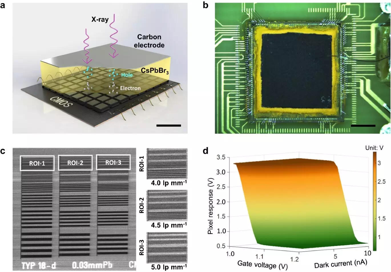
The research team successfully developed a direct-conversion X-ray detector using a 300 μm thick inorganic CsPbBr3 perovskite film printed on a dedicated CMOS pixel array. The results of the study demonstrated that the thick CsPbBr3 film exhibited a high μτ product, high X-ray detection sensitivity, and a low dose detection limit, making it a promising candidate for high-performance X-ray imaging.
The Impressive Imaging Performance
Experimental X-ray 2D imaging results showed that the perovskite CMOS detector achieved exceptional spatial resolution and low-dose imaging performance. With a spatial resolution of 5.0 lp mm-1 and a low dose of 260 nGy, the detector outperformed traditional imaging systems. Additionally, 3D CT imaging was validated with the proposed detector, showcasing its fast signal readout speed of 300 fps.
The study highlights the immense potential of lead halide perovskites in transforming the development of state-of-the-art X-ray detectors. With enhanced spatial resolution, readout speed, and low-dose detection efficiency, the perovskite X-ray CMOS detector opens up new possibilities in the field of medical imaging.
Leave a Reply