
In a groundbreaking study published in the journal Optica, researchers at HHMI’s Janelia Research Campus have revolutionized the field of microscopy by adapting techniques traditionally used in astronomy to improve the clarity and sharpness of images. This innovative approach has the potential to provide biologists with a more cost-effective and efficient way to capture detailed images of biological samples.
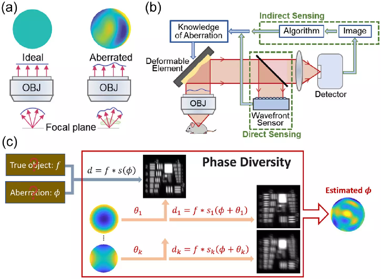
Astronomers have long been utilizing techniques to enhance the images captured by their telescopes, taking into account the distortions caused by the atmosphere to produce clearer and sharper pictures of distant galaxies. In a similar fashion, microscopists have been exploring the application of adaptive optics methods to correct for aberrations in microscopy images of thick biological samples. However, traditional adaptive optics methods have been deemed complex, expensive, and slow, limiting their accessibility to many research labs.
To address the limitations of conventional adaptive optics techniques, the research team at Janelia Research Campus turned their focus to phase diversity methods, a class of techniques widely used in astronomy but relatively new to the life sciences. By incorporating additional images with known aberrations into a blurry image with unknown aberrations, phase diversity methods provide the necessary information to unblur the original image, without requiring significant alterations to the imaging system.
In order to implement the novel phase diversity method in microscopy, the team first adapted the astronomy algorithm for use in biological imaging and verified its effectiveness through simulations. Subsequently, they constructed a microscope equipped with a deformable mirror and two additional lenses, minor modifications that introduce the known aberrations into the system. Additionally, the team enhanced the software used to execute the phase diversity correction, streamlining the calibration process of the deformable mirror by a significant margin.
Through rigorous testing, the research team demonstrated the capability of the new method to sense and correct randomly generated aberrations, resulting in clearer images of fluorescent beads and fixed cells. Moving forward, the team plans to evaluate the method on real-world samples, including living cells and tissues, while exploring its application in more complex microscopy setups. Furthermore, they aim to streamline the method, making it more automated and user-friendly to broaden its accessibility to researchers across various disciplines.
The introduction of phase diversity techniques from astronomy into the realm of microscopy represents a significant advancement in the field, offering a faster, cheaper, and more user-friendly approach to improving image quality. By adapting these innovative methods, researchers hope to empower biologists with the tools necessary to delve deeper into the complexities of biological samples, ultimately enhancing our understanding of the microscopic world.
Leave a Reply