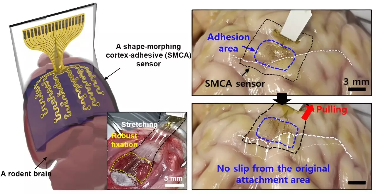
As the field of neurology continues to evolve, innovative technologies are emerging to tackle complex neurological disorders. One of the most promising developments is the use of transcranial focused ultrasound (TFUS), a non-invasive technique that utilizes high-frequency sound waves to stimulate specific regions of the brain. This method shows particular promise for conditions such as drug-resistant epilepsy and other movement disorders characterized by tremors. Recently, a multidisciplinary research team led by experts from Sungkyunkwan University and the Korea Institute of Science and Technology unveiled an advanced sensor designed to enhance the efficacy of TFUS.
Historically, sensors aimed at monitoring brain activity posed significant challenges, particularly when it came to achieving accurate and reliable measurements. The intricate folds and contours of the brain often meant that conventional brain sensors could not maintain solid contact, leading to inconsistent readings and potentially misleading diagnoses. Donghee Son, a principal investigator in the study, highlights a critical drawback of earlier devices, stating, “Previous brain sensors struggled with curvature and motion, which directly impacted the accuracy of brain signal readings.” Even groundbreaking earlier technologies that reduced the sensor’s thickness failed to achieve suitable adhesion on notably curved surfaces.
These limitations have significant implications for clinical practice, as they restrict the ability to monitor brain activity continuously and accurately, especially in patients with highly variable conditions. The need for a more adept solution was imperative, particularly for personalized treatment plans in epilepsy, where the variability and intricacies of brain function in each individual can make standard treatments less effective.
In efforts to address these challenges, the collaborative team embarked on developing a novel sensor designed to conform closely to the brain’s surface and remain secure regardless of the curvature. The newly created ElectroCorticography (ECoG) sensor excels in maintaining stable, long-term adhesion, which is crucial for chronic conditions that demand real-time monitoring. With this new sensor, the precision of neural signal measurement is significantly enhanced, allowing for effective real-time analysis and control of seizures.
The new design features three distinct layers: a hydrogel that bonds chemically and physically with brain tissue, a self-healing polymer that adapts to surface contours, and a stretchable layer housing gold electrodes. Son explains, “The melding of adhesive and shape-morphing technologies enables our sensor to achieve strong, stable attachment, which drastically reduces external noise—an essential factor in enhancing treatment effectiveness through low-intensity ultrasound.”
Implications for Personalized Medicine
The significance of this advancement cannot be overstated. Traditional sensors have struggled with noise from ultrasound vibrations, which made it nearly impossible to monitor brain activity accurately while using ultrasound treatment. The new sensor integrates a means of minimizing these vibrations, thereby facilitating the development of tailored treatment regimens for epilepsy and other neurological disorders.
Personalized medical solutions are increasingly recognized as key to treating complex conditions effectively. Being able to monitor brain waves in real time, while simultaneously delivering targeted ultrasound stimulation, opens doors for neurological treatments that are adaptable to each patient’s unique condition. Son emphasizes, “Our sensor’s ability to effectively reduce noise enhances the potential for personalized treatment strategies, which is a significant advancement in neurology.”
Future Directions and Clinical Applications
The initial findings have been promising, demonstrating the sensor’s effectiveness in living rodent subjects, where precise measurements of brain signals and seizure control were successfully achieved. The researchers aim to expand upon this work, with plans for a high-density electrode array that would further improve the resolution of brain signal mapping. “While our current model is equipped with 16 electrode channels, scaling up will be essential for achieving higher-resolution brain signal analysis,” notes Son.
The ultimate goal of this research extends beyond epilepsy treatment. Should clinical trials confirm the efficacy and safety of this technology in human subjects, the sensor could become a pivotal tool for diagnosing and treating a variety of neurological conditions. Furthermore, the innovations in sensor technology could catalyze advancements in adjacent fields, including the development of more effective prosthetic technologies, proving that the intersection of engineering and neurology may well shape the future of healthcare.
The collaboration of technology and medicine has delivered a groundbreaking tool capable of changing the landscape of neurological treatment, heralding unprecedented possibilities for personalized patient care and enhanced therapeutic strategies. With strong initial results and ambitious future plans, the journey from laboratory success to clinical application stands to revolutionize the approach to managing neurological disorders.


Leave a Reply