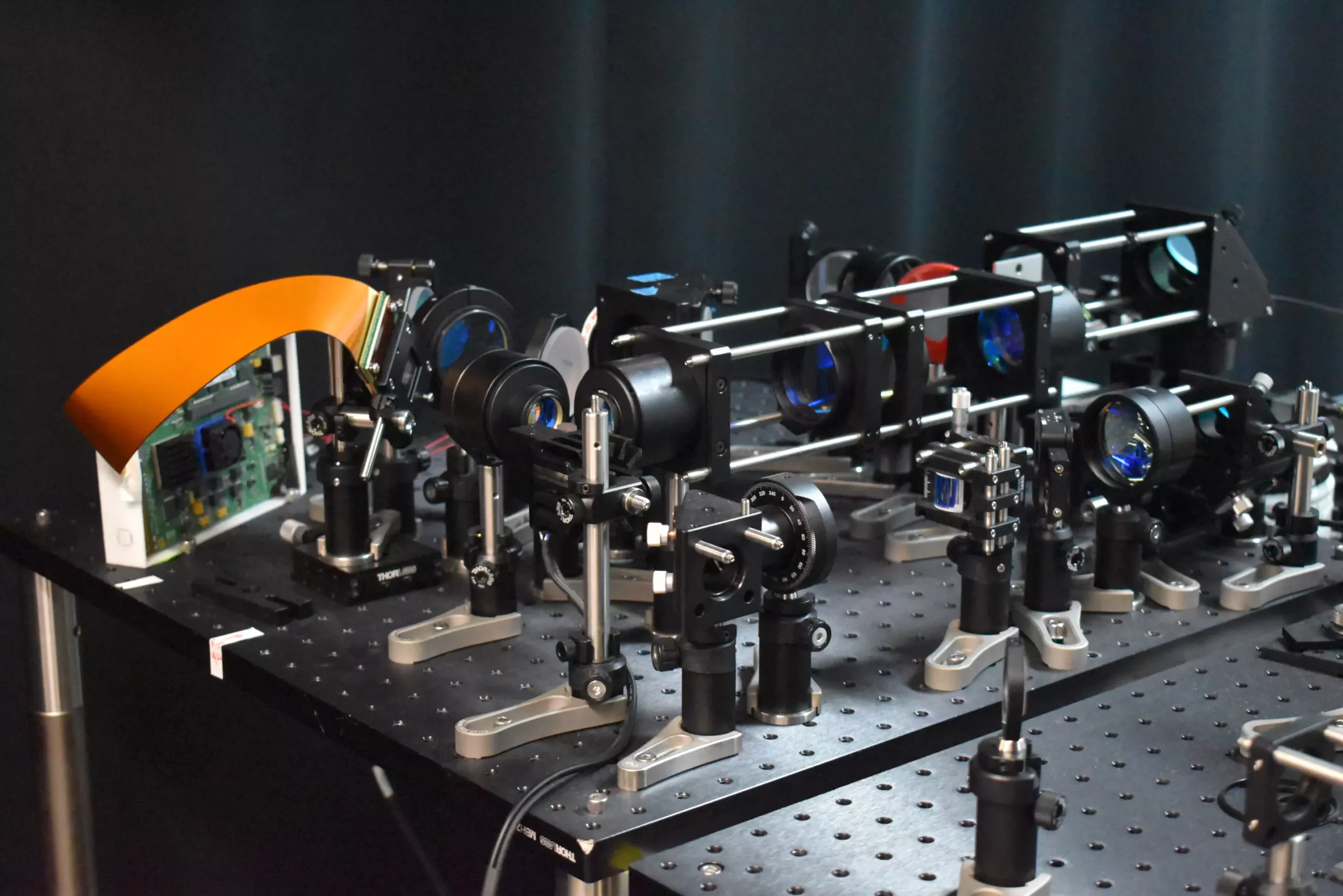
Researchers continue to push the boundaries of neuroscience with the development of a new two-photon fluorescence microscope. This innovative tool promises high-speed imaging of neural activity at cellular resolution, allowing for a more in-depth understanding of how neurons communicate in real time. Led by Weijian Yang from the University of California, Davis, the research team behind this groundbreaking technology aims to revolutionize the field of neuroscience.
Enhanced Imaging Capabilities
Traditionally, two-photon microscopy has provided detailed images of neural activity but at a slow pace and with the potential for damaging brain tissue. However, the new approach described by the researchers introduces a novel adaptive sampling scheme that replaces traditional point illumination with line illumination. This technique enables in vivo imaging of neuronal activity in a mouse cortex at speeds ten times faster than conventional two-photon microscopy, all while significantly reducing the laser power on the brain.
The implications of this breakthrough extend beyond fundamental brain functions like learning and memory. The new two-photon fluorescence microscope could play a crucial role in studying the pathology of neurological diseases at their earliest stages. By observing neural activity in real time, researchers hope to gain a better understanding of diseases such as Alzheimer’s, Parkinson’s, and epilepsy. This technology could potentially lead to more effective treatments and interventions for these conditions.
The researchers implemented an adaptive sampling strategy utilizing a digital micromirror device (DMD) to target active neurons in the brain tissue. This strategy allows for a larger area to be imaged at once, speeding up the imaging process and reducing the risk of damage to brain tissue. By dynamically shaping and steering the light beam with thousands of tiny mirrors, the researchers achieved precise targeting of active neurons, improving the overall efficiency of the imaging process.
The capabilities of the new microscope were demonstrated through imaging of calcium signals in living mouse brain tissue at a speed of 198 Hz, showcasing its superiority over traditional imaging methods. The adaptive line-excitation technique, coupled with advanced computational algorithms, enables the isolation of individual neuronal activity, a vital aspect for decoding complex neural interactions and unraveling the brain’s functional architecture.
Looking ahead, the researchers aim to integrate voltage imaging capabilities into the microscope for a rapid readout of neural activity. They also plan to utilize the technology in real neuroscience applications, such as observing neural activity during learning processes and studying brain activity in disease states. Additionally, efforts will be made to enhance the user-friendliness of the microscope and reduce its size to broaden its utility in neuroscience research.
The development of the new two-photon fluorescence microscope represents a significant step forward in the field of neuroscience. With its enhanced imaging capabilities, targeted sampling technique, and potential applications in understanding neurological diseases, this technology holds great promise for advancing our knowledge of the brain’s intricate workings. By continuing to refine and expand upon this innovative tool, researchers may unlock new insights into neural processes and pave the way for transformative discoveries in neuroscience.


Leave a Reply