
Nonlinear light microscopy has opened up new possibilities in observing and interpreting complex biological processes. However, the use of intense light in this technique raises concerns about the potential damage it can cause to living matter. Despite its benefits, the mechanism behind the irreversible perturbation of cellular processes by intense light remains a significant gap in our understanding.
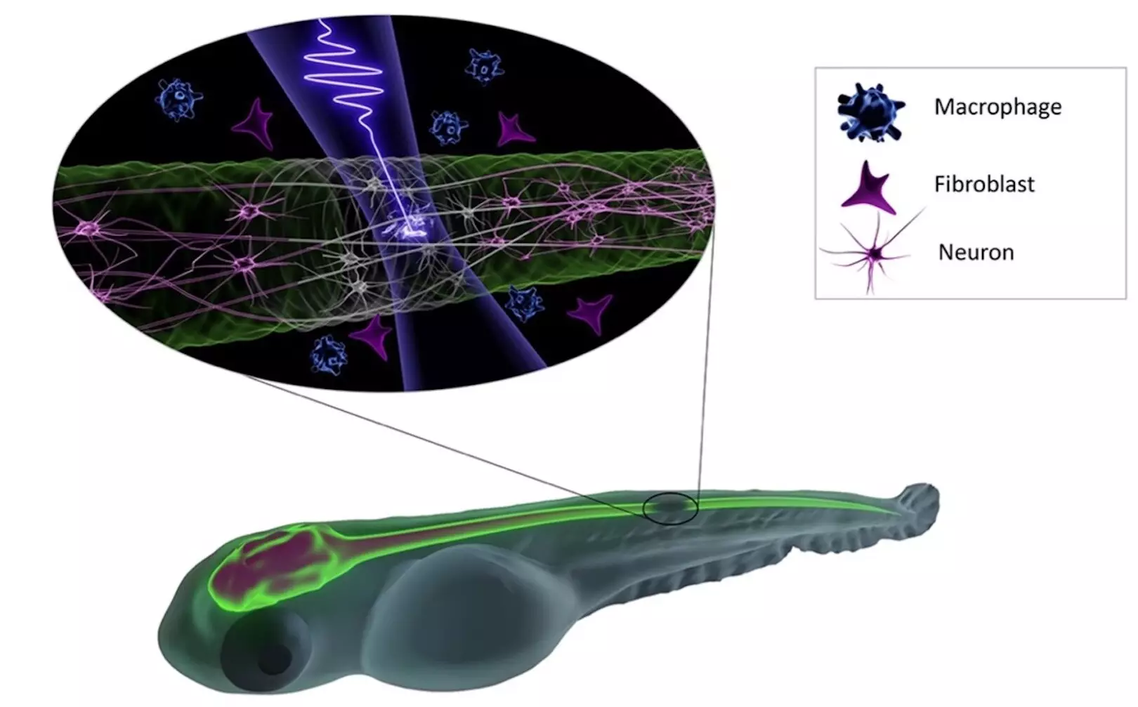
To address this issue, the research teams led by Hanieh Fattahi and Daniel Wehner at the Max Planck Institute for the Science of Light and Max-Planck-Zentrum für Physik und Medizin collaborated to investigate the conditions under which intense pulsed lasers can be utilized in vivo without harming the organism. By using zebrafish as a model organism, the international team from Erlangen explored the mechanisms of photodamage in deep tissue at a cellular level induced by femtosecond excitation pulses.
Key Findings on Photodamage in Zebrafish
The study, published in Communications Physics, revealed that damage to the central nervous system of zebrafish occurred abruptly when exposed to femtosecond pulses at 1030 nm, reaching peak intensities necessary for low-density plasma formation. The research demonstrated that noninvasive increase in imaging dwell time and photon flux during irradiation at 1030 nm was possible as long as the peak intensity stayed below the low-plasma density threshold. These findings have significant implications for nonlinear label-free microscopy and can advance deep tissue imaging techniques.
The development of innovative microscopy techniques, such as femtosecond fieldoscopy, holds promise for capturing high spatial resolution, label-free images with attosecond temporal resolution. According to Fattahi, the technique could revolutionize the field of microscopy by offering precise manipulations of the central nervous system and opening up new possibilities for in vivo applications. The collaboration between the fields of physics and biology not only shed light on the impact of intense light on living matter but also paves the way for future advancements in deep tissue imaging and innovative microscopy techniques.
The study on the effects of femtosecond excitation pulses on deep tissue imaging in zebrafish provides valuable insights into the mechanisms of photodamage and opens up new possibilities for noninvasive imaging techniques. By understanding the conditions under which intense pulsed lasers can be used without causing harm, researchers can further advance the field of nonlinear light microscopy and explore new avenues for in vivo applications in biology and medicine.
Leave a Reply